Advances in Animal and Veterinary Sciences
Research Article
Advances in Animal and Veterinary Sciences 1 (1): 28–31.Incidence of Mycobacterium avium subspecies paratuberculosis in Mehsana Breed of Goats from North Gujarat using Multiple Tests
Khushboo Singh1, Bharat Singh Chandel1, Abidali I Dadawala1, Shoor Vir Singh2*, , Harshad C. Chauhan1, Brajesh Singh2, Narottam Das Agrawal2, Saurabh Gupta2, Kundan Kumar Chaubey2
- Department of Veterinary Microbiology, College of Veterinary Science & Animal Husbandry, Sardarkrushinagar Dantiwada Agricultural University, Sardarkrushinagar– 385506, Gujarat, India
- Microbiology Laboratory, Animal Health Division, Central Institute for Research on Goats, Makhdoom, PO – Farah, Dist.– Mathura, 281 122, Uttar Pradesh, India
*Corresponding author:shoorvir.singh@gmail.com; shoorvir_singh@rediffmail.com
ARTICLE CITATION:
Singh K, Chandel BS, Dadawala AI, Singh SV, Chauhan HC, Singh B, Agrawal ND, Gupta S, Chaubey KK (2013). Incidence of Mycobacterium avium subspecies paratuberculosis in Mehsana breed of goats from south Gujarat using multiple tests. Adv. Anim. Vet. Sci. 1 (1): 28–31.
Received: 2013–04–03, Revised: 2013–04–11, Accepted: 2013–04–12
The electronic version of this article is the complete one and can be found online at
(
http://www.nexusacademicpublishers.com/table_contents_detail/4/29/html
)
which permits unrestricted use, distribution, and reproduction in any medium, provided the original work is properly cited
ABSTRACT
The study on incidence of Johne’s disease in goat envisaged detection of MAP antibodies, clinico–pathological observations in naturally infected goats, detection of MAP in feces and blood. More than 70 percent goats showed the clinical symptoms of loss of appetite, dullness, emaciation, loss of hairs (alopecia), pasty or loose feces suggestive of Johne’s disease. A total of 200 serum samples were screened for detecting presence of MAP antibodies using indigenous ELISA (i–ELISA) kit yielding 63.5% seroprevalence. Samples viz., feces and rectal pinch were collected from 50 goats (strong reactors in ELISA) were screened for MAP bacilli by Z–N staining. Out of these, 14 (28.0%) showed the presence of acid fast bacilli indistinguishable to MAP, whereas none of the goat was positive in rectal pinch smear. This study was further followed by detection of MAP genome in feces of direct microscopy positive animals and blood from strong positive ELISA reactors by IS900 PCR. Of 14 fecal and 50 blood samples, 1 (7.14%) and 6 (12.0%) were positive in fecal and blood PCR, respectively.
INTRODUCTION
Johne’s disease is a chronic granulomatous enteropathy of domestic and wild ruminants, caused by Mycobacterium avium subsp. paratuberculosis (MAP) is of major concern in developed countries (Singh et al., 2013a and 2013b). Concerns have been raised about the apparent increased global prevalence of MAP, leading to increased economic costs. The ability to detect MAP accurately and rapidly is an integral part of herd management. However, detection and control of this bacterium is complicated due to its slow division time and its ability to persist in the environment (Singh et al., 2013b). Effective detection of subclinical cases of bovine Johne’s disease is a critical step in the reduction of disease prevalence in dairy herds (Eamens et al., 2000). JD has not received due priority in India. This is primarily due to lack of indigenous diagnostic kits and reagents. MAP infecting goats has been genotyped as ‘Bison type’ in North India (Sevilla et al., 2005, Singh et al., 2010a). Using ‘Bison type’ genotype as source of antigen, indigenous ELISA kit was developed for goats (Kumar et al., 2006; Singh et al., 2007a) and used to screen goats in the present study. The JD is endemic in farm and farmer’s goatherds in India (Singh and Vihan, 2004, Singh et al., 2007b, Kumar et al., 2007). Singh et al. (1998) reported higher prevalence of JD in herds located in semi arid region as compared to arid region. However, information is limited on the prevalence of JD in Gujarat. This study aimed to ascertain the incidence of paratuberculosis in Mehsana goats from organized farm of Sheep and Goat Research Station (SGRS), Gujarat in the year 2011 by means of direct microscopy, IS900 PCR and ELISA kit.
MATERIALS AND METHODS
Animals and samples (feces, blood and serum)
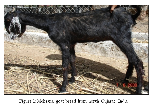
Incidence of paratuberculosis was studied in Mehsana goats from organized goatherd located at Sheep and Goat Research Station (SGRS), Gujarat in the year 2011. The animals were examined clinically for symptoms resembling to JD (Figure 1). Body conditions viz., hair coat, skin texture etc were also observed and recorded.
A total of 200 sera were collected. Apart from this, a total of 50 fecal samples and 50 blood samples were also collected from goats having strong positive ELISA titer. Forteen rectal pinch smears was collected from the animals that were found positive in direct microscopy. Samples were screened by ELISA, direct microscopy, fecal and blood IS900 PCR.
Screening of Serum Samples of Goats by Indigenous ELISA Kit
Indigenous ELISA kit developed at Central Institute of Research on Goats (CIRG), Makhdoom was used for screening of 200 serum samples, strictly as per the protocol outlined in the user’s manual supplied with the kit. Since JD is a spectral disease, instead of single point cutoff, OD values of ELISA were used to calculate S/P ratios (Collins 2002). Animals in positive and strong positive category were considered as positive.
Direct Microscopy
About 2–3 grams of fecal sample was collected in polythene bag from goats having strong positive ELISA titer, directly from rectum. Samples were homogenized and concentrated by centrifugation at at 4000 rpm for 45 min at room temperature. Supernatant was discarded and from middle layer smears were prepared, stained by Ziehl Neelsen (ZN) staining and were examined under oil immersion (100X) for presence of acid–fast bacilli (AFB) indistinguishable to MAP.
Rectal Pinch Examination
Rectal pinch smear examination was performed only in animals which were found positive for MAP bacilli in direct fecal microscopy. The rectal pinch was collected with a sterile artificial insemination sheath. Smears were prepared on grease free slide, heat fixed and stained by Ziehl–Neelsen (ZN) staining. On the basis of presence of pink colored rod shaped bacilli, the samples were recorded as positive.
IS900 PCR
Forteen fecal samples and 50 blood samples were processed for DNA isolation as per Van Embden et al. (1993) and Singh et al. (2010b). The samples were screened for the presence of MAP genome in the positive fecal samples and blood sample using IS900 PCR on extracted DNA to obtain the frequency of distribution of MAP in Mehsana goats maintained at SGRS.
DNA samples were amplified using specific IS900 (P90 and P91) primers (Millar et al., 1996). Briefly, in a volume of 12.5 μl of 2X master mix, 1 μl forward primer (10 pmole/μl) and 1μl reverse primer (10 pmole/μl), 5.5 μl of nuclease free water and 5 μl of template DNA was added (total volume 25 μl). Total of 37 cycles were performed in a thermocycler (MJ research) for complete amplification reaction. Thermal cycling conditions were: initial denaturation at 94 oC for 3 min (1 cycle), denaturation at 94 o for 30 s, annealing at 62 o for 15 s, extension at 72 o for 1 min (37 cycles) and final extension at 72 o for 10 min. Presence and yield of specific 413bp product was analyzed by 1.5% agarose ethidium bromide gel electrophoresis.
RESULTS AND DISCUSSION
In the present study incidence of Johne’s disease was estimated in Mehsana goats maintained at Sheep and Goat Research Station, Sardarkrushinagar, Gujarat. More than 70 per cent goats showed the clinical signs resembling to Johne’s disease, which included loss of appetite, dullness, emaciation, rough hair coat, loss of hairs (alopecia), pasty or loose fecal, with tail hairs sticked together. Paliwal and Rajya (1982) and Barad (2009) in their study also observed the similar clinical conditions in goats in Uttar Pradesh and Gujarat, respectively. However, Molina et al. (1991) observed sub–mandibular oedema and cachexia in goats which were clinically affected from Johne's disease.
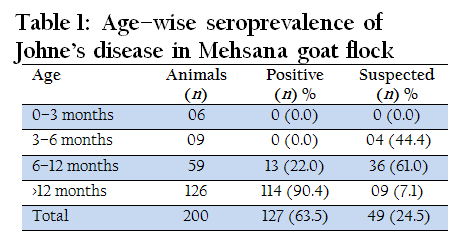
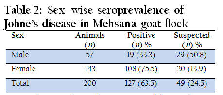
Serological surveys are good methods for assessing the prevalence of disease in particular disease in particular species of animals. Though, specificity is less than culture, serology provides a cost effective alternative to organism detection based diagnostic tests for Johne’s disease. ELISA is the most sensitive and specific test for detection of serum antibodies against M. paratuberculosis (OIE manual, 2000) and is best used as a herd or flock screening tool. A total of 200 serum were screened and of these 127 (63.5 %) were found to be positive for the presence of MAP antibodies (Table 1 and Table 2). Similar to present findings a higher prevalence of 55.1 per cent has also been reported by Vohra et al. (2008) in goats of CIRG, Makhdoom. Singh et al. (2007a) and Singh et al. (2007b) reported a lower sero–prevalence rate of 19.6–31.8 per cent and 30.9–50.0 per cent in goats of Western Rajasthan and Uttar Pradesh, respectively. Goswami et al. (2000) has also reported a very low sero–prevalence of 13.5 per cent in an organized farm of Pashmina goats. The possible reason for the higher rate of sero–prevalence recorded during present study may be the higher incidence of the disease in the herd and goats were suffering from clinical JD since longer, which would have resulted into sero–conversion in the affected goats.
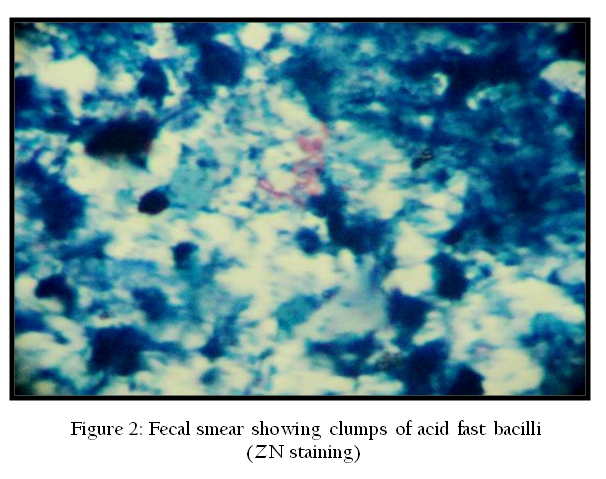
A total of 50 fecal samples from the animals which were suffering from diarrhea or pasty fecal and had strong positive ELISA titer were microscopically examined for detecting MAP bacilli in the fecal using Ziehl–Neelsen's staining and 14 (28.0) per cent were found to be positive (Figure 2). Barad (2009) reported very low prevalence of MAP shedders 8.93 per cent out of 56 goats in Gujarat. The reason of high incidence of the MAP shedders might be that in the present study, samples were collected from animals more than 6 months of age suffering from clinical JD. This might have contributed in the higher prevalence of MAP in fecal sample. Also, the animals are maintained completely on grazing, the fecal might have contaminated the environment where all the goats irrespective of the age group are kept together and hence acting as a source of increasing the uptake of infection. 14 goats found positive for MAP bacilli in fecal sample, were screened for presence of MAP bacilli in rectal pinch smear. None of the goat was found to be positive which collaborates with the findings of Barad (2009). It is described in Ohio State University Fact Sheet for Johne’s disease in sheep and goats that the Smears of rectal mucosa biopsies are difficult to get and may not be very helpful in detection of JD because the disease process is less likely to involve the rectum in sheep and goats. (http://ohioline.osu.edu/vme–fact/0003.html).
In accordance with a technique (van Embden et al., 1993) common extraction and enhancement technique was followed. Goats were screened for the presence of MAP genome in the positive fecal samples using IS900 PCR on DNA extracted from fecal to obtain the frequency of distribution of MAP infection in Mehsana goats maintained at SGRS. Out of 14 fecal samples obtained from the goats having history of recurrent diarrhea or pasty fecal, 1 (7.14) per cent was found positive (Figure 3). In contrast to present findings Barad (2009) found 12.5 per cent fecal PCR positive cases out of 40 JD suspected goats in Gujarat. The difference in the findings might be due to the intermittent shedding of the MAP bacilli in the fecal or variation in the genotype of MAP or the presence of PCR inhibitors.

Detection of MAP by IS900 PCR in fecal samples though rapid but is low throughput in kids as MAP shedding at early to sub–clinical stage is rare or intermittent and also due to the presence of PCR inhibitors (Harris and Barletta, 2001). So, the present study also aimed to detect MAP genome in blood using IS900 PCR on DNA extracted from blood. A total of 50 goats, which were found strongly positive reactors in ELISA were screened to obtain the frequency of distribution of MAP. Of 50 blood samples screened 6 (12.0) per cent goats were found positive (Figure 3). In contrast to the present findings Singh et al. (2010b) reported higher prevalence of 77.5 per cent of MAP genome in blood PCR in goats maintained at CIRG, Makhdoom. They concluded that possible factor for increased detection was associated with improved optimization of blood PCR in naturally infected goats and higher level of infections. Coelho et al. (2008) also estimated the higher prevalence of 20.7 per cent of MAP genome in pooled samples from apparently healthy and 16.7 per cent in blood PCR in pooled samples from suspected sheep blood. The reason for the low positive finding in the present study might be the stage or the level of the infection or storage conditions of samples or due to less severe extra intestinal infection.
CONCLUSION
In the present study, it has been found that JD is endemic in Mehsana goat herd at SGRS farm, Gujarat. More than 70 per cent affected goats showed loss of appetite, dullness, emaciation, alopecia, pasty or loose fecal. Overall sero–prevelance of MAP antibodies recorded was 63.5% (127 out of 200). Of 50 strong positive ELISA reactors, 14 (28.0%) goats were found positive in direct fecal microscopy. All goats were found negative for MAP bacilli in rectal pinch smear examination. Overall MAP bacterimia in fecal and blood was 7.14 and 12.0 % from 14 direct microscopy positive goats and 50 strongly ELISA positive reactors, respectively. On the basis of present study there is need for immediate implementation of control programmes for JD at National level.
ACKNOWLEDGMENT
Authors are thankful to Department of Microbiology, College of Veterinary Science and Head, GHD and Director, CIRG for extending necessary facilities for the work.
REFERENCES
Barad DB (2009). Comparative evaluation of molecular, serological and conventional diagnostic methods for Johne's disease in goats. M.V.Sc. thesis, Sardarkrushinagar Dantiwada Agriculture University. Sardarkrushinagar. Gujarat.
Coelho AC, Pinto ML, Coelho AM, Rodrigues J and Juste R (2008). Estimation of the prevalence of Mycobacterium avium subsp. paratuberculosis by PCR in sheep blood. Small Rumin. Res. 76(3): 201 - 206.
http://dx.doi.org/10.1016/j.smallrumres.2007.12.003
Collins MT (2002). Interpretation of a commercial bovine paratuberculosis enzyme-linked immunosorbent assay by using likelihood ratios. Clin. Diagn. Lab. Immunol. 9(6): 1367 - 1371.
PMid:12414776 PMCid:PMC130105
Eamens GJ, Whittington RJ, Marsh IB, Turner JJ, Saunders V, Kemsley PD and Rayward D (2000). Comparative sensitivity of various fecal culture methods and ELISA in dairy cattle herds with endemic Johne's disease. Vet. Microbiol. 77(3-4): 357 - 368.
http://dx.doi.org/10.1016/S0378-1135(00)00321-7
Goswami TK, Tiwari V, Gupta A, Mall R and Ram GC (2000). Seroprevalence of Mycobacterium paratuberculosis in an organized Pashmina goat farm. Indian. J. Comp. Microbiol. Immunol. Infect. Dis. 21(2): 132 - 135.
Harris NB and Barletta RA (2001). Mycobacterium avium subsp. paratuberculosis in veterinary medicine. Clin. Microbiol. Rev. 14(3): 489 - 512.
http://dx.doi.org/10.1128/CMR.14.3.489-512.2001
PMid:11432810 PMCid:PMC88986
Kumar P, Bhatiya AK and Singh SV (2006). Evaluation of efficacy of the species specific antigens in the diagnosis of ovine and caprine Paratuberculosis using plate ELISA. J. Immunol. Immunopath. 8: 48 - 53.
Kumar P, Singh SV, Bhatiya AK, Sevilla I, Singh AV, Whittington RJ, Juste RA, Gupta VK, Singh PK, Sohal JS and Vihan VS (2007). Juvenile capri-paratuberculosis (JCP) in India; Incidence and characterization by six diagnostic tests. Small Rumin. Res. 73(1-3): 45 - 53.
http://dx.doi.org/10.1016/j.smallrumres.2006.10.023
Millar D, Ford J, Sanderson J, Withey S, Tizard M, Doran T, Hermon-Taylor J (1996). IS900 PCR to detect Mycobacterium paratuberculosis in retail supplies of whole pasteurized cows milk in England and Wales. Appl. Environ. Microbiol. 62(9): 3446 - 3452. PMid:8795236 PMCid:PMC168142
Molina A, Morere L and Lianes D (1991). Enzyme-linked immunosorbent assay for detection of antibodies against Mycobacterium paratuberculosis in goats. Am. J Vet. Res., 52(6): 863 - 868.
PMid:1883088
OIE Manual (2000). Johne's disease. pp. 30 - 35.
Paliwal OP and Rajya BS (1982). Evaluation of paratuberculosis in goats: Pathomorphological studies. Indian J. Vet. Path. 6: 29 - 34.
Sevilla I, Singh SV, Garrido JM, Aduriz G, Rodriguez S, Geijo MV, R.J. Whittington, V. Saunders, R.H. Whitlock and R.A. Juste (2005). PCR-REA genotype paratuberculosis strains isolated from different host and species and geographic locations. Rev-Off. Int. Epizoot. 24(3): 1061 - 6.
Singh AV, Singh SV, Singh PK and Sohal JS (2010a). Genotype diversity in Indian isolates of Mycobacterium avium subspecies paratuberculosis recovered from domestic and wild ruminants from different agro-climatic regions. Comp. Immunol. Microbiol. Infect. Dis. 33(6): e127 - 131.
http://dx.doi.org/10.1016/j.cimid.2010.08.001
PMid:20832117
Singh N, Vihan VS, Singh SV and Gupta VK (1998). Prevalence of Johne's disease in organized goatherds. Indian. J. Anim. Sci. 68: 41 - 42.
Singh PK, Singh SV, Kumar, H, Sohal JS and Singh AV (2010b). Diagnostic Application of IS900 PCR Using Blood as a Source Sample for the Detection of Mycobacterium avium subspecies paratuberculosis in Early and Subclinical Cases of Caprine Paratuberculosis. Vet. Med. International. doi:10.4061/2010/748621.
http://dx.doi.org/10.4061/2010/748621
Singh SV and Vihan VS (2004). Incidence of Mycobacterium avium subspecies paratuberculosis in clinically suspected small ruminants. Small Rumin. Res. 34: 231 - 235.
http://dx.doi.org/10.1016/j.smallrumres.2003.12.002
Singh SV, Singh AV, Singh PK, Sohal JS and Singh NP (2007a). Evaluation of an indigenous Elisa for diagnosis of Johne's disease and its comparison with commercial kits. Indian J. Microbiol. 47(3): 251 - 258.
http://dx.doi.org/10.1007/s12088-007-0046-2
PMid:23100673 PMCid:PMC3450340
Singh SV, Singh AV, Singh PK, Sohal JS, Vikram Y and Kharche SD (2007b). Sero-prevalence of Mycobacterium avium subspecies paratuberculosis infection in Barbari and Sirohi breeds of goats in their respective home tracts in semi-arid regions of North India. Haryana Vet. 46: 104 - 106.
Singh SV, Dhama K, Chaubey KK, Kumar N, Singh PK, Sohal JS, Gupta S, Singh AV, Verma AK, Tiwari R, Mahima Chakraborty S and Deb R (2013a). Impact of Host Genetics on susceptibility and resistance to Mycobacterium avium subspecies Paratuberculosis in domestic ruminants. Pak. J. Biol. Sci. 16(6): 251 - 266.
http://dx.doi.org/10.3923/pjbs.2013.251.266
PMid:24498788
Singh SV, Singh AV, Kumar A, Singh PK, Deb R, Verma AK, Kumar A, Tiwari R, Chakraborty S and Dhama K (2013b). Survival mechanisms of Mycobacterium avium subspecies paratuberculosis within host species and in the environment- A review. Natural Science (article in press).
http://dx.doi.org/10.4236/ns.2013.56088
Stott AW, Jones GM, Humphry RW and Gunn GJ (2005). Financial incentive to control paratuberculosis (Johne's disease) on dairy farms in the United Kingdom. Vet. Rec. 156(26): 825 - 831.
PMid:15980134
Van Embden JDA, Cave D, Crawford JT, Dale JW, Eisenach KD and Gicquel B (1993). Strain identification of Mycobacterium tuberculosis by DNA fingerprinting: recommendations for a standardized methodology. J Clin. Microbiol. 31(2): 406 - 9.
PMid:8381814 PMCid:PMC262774
Vohra J, Singh SV, Singh AV, Singh PK and Sohal JS (2008). Comparative distribution of Mycobacterium avium subspecies paratuberculosis in the target and non-target tissues of goats and sheep population in India. Indian J. Anim. Sci. 78(4): 342 - 347.