Advances in Animal and Veterinary Sciences
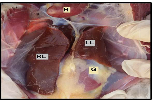
The liver in mallard duck. Showing the left lobe (LL), right lobe (RL) of the liver, heart (H), gizzard (G).
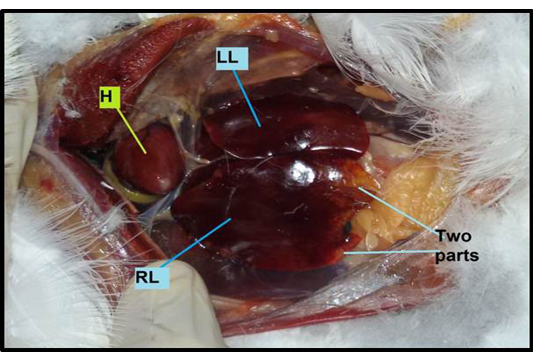
The liver in gull. Showing the left lobe (LL), right lobe (RL) which contains two parts, a presence Heart (H).
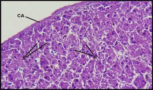
Histological section of the liver in gull: Showing the thick capsule (CA), hepatocyte (H), sinusoids (S). (H&E 40X).
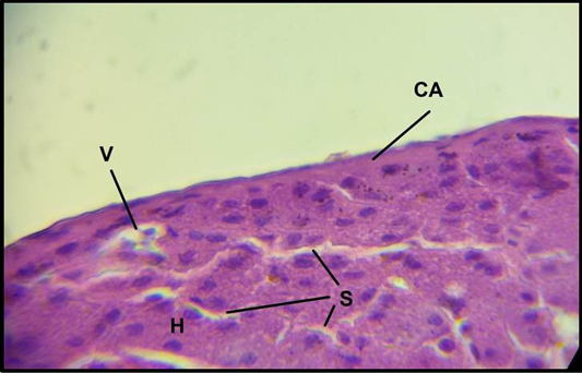
Histologicalsection of the liver in mallard duck s: Showing the capsule (CA), hepatocyte (H), sinusoids (S), vein (V). (H&E 100X).
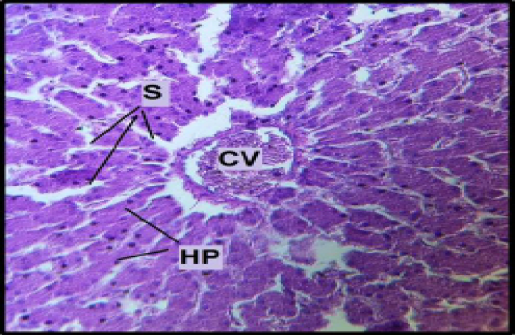
Histological section of the liver in mallard duck: Showing Central vein (CV), Hepatocyte Plates (HP), Sinusoids (S). (H&E 40X).
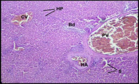
Histological section of the liver in gull : Showing Central vein (CV), Hepatocyte plates (HP), Sinusoids (S) Portal vein (PV), Hepatic artery (HA), Bile duct(Bd). (H&E 20X).
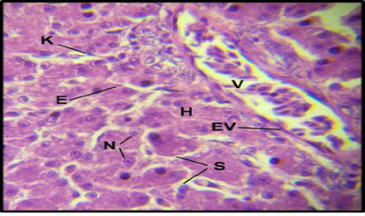
Histological section of the liver in mallard duck: Showing Vein (V), Hepatocyte (H), Sinusoids (S). Nucleus of hepatocyte (N), kupffer cell (K), Endothelial cell of sinusoid (E), Endothelial cells of Vein (EV). (H&E 100X).
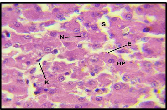
Histological section of the liver in gull: Showing Hepatocyte Plates (HP), sinusoids (S), Nucleus of hepatocyte (N), kupffer cell (K), Endothelial cell of sinusoid (E). (H&E 100X).
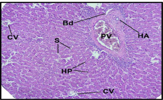
Histological section of the liver in mallard duck : Showing the portal triad. Portal vein (PV), Hepatic artery (HA), Bile duct (Bd), Central vein (CV) Sinusoids (S), Hepatic plate (HP)(H&E 20X).
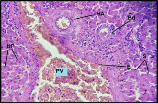
Histological section of the liver in gull: Showing the portal triad. Portal vein (PV), Hepatic artery (HA), Bile duct (Bd) Sinusoids (S), Hepatic plate (HP), Endothelial cells of portal vein (E). (H&E 40X).


