Advances in Animal and Veterinary Sciences
Research Article
Adv. Anim. Vet. Sci. 4(12): 642-647
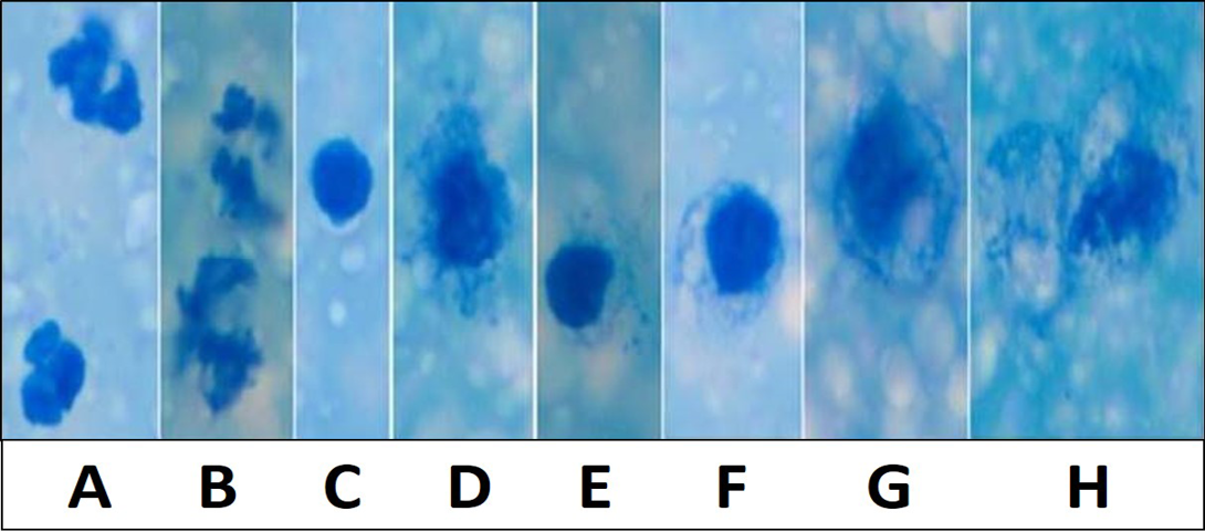
Figure 1
Various inflammatory and secretory cells Newman’s stain X 1000: A) Polymorphonuclear cells/ Neutrophils; B) Degenerating neutrophils; C) Lymphocytes; D and E) Macrophages with irregular cell membrane and basophilic engulfed material; F and G) Desquamated secretory glandular epithelial cells; H) Large desquamated secretory glandular epithelial cell with granulesMicroscopically milk showing occasional inflammatory cells in Newman’s stain X 200
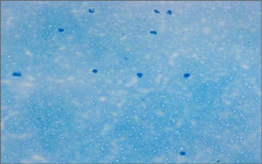
Figure 2
Microscopically mastitis infected milk showing abundant inflammatory cells in Newman’s stain X 200
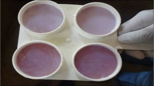
Figure 3
Detection of SCM by CMT test
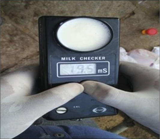
Figure 4
Detection of mastitis by EC test using milk checker instrument


