Advances in Animal and Veterinary Sciences
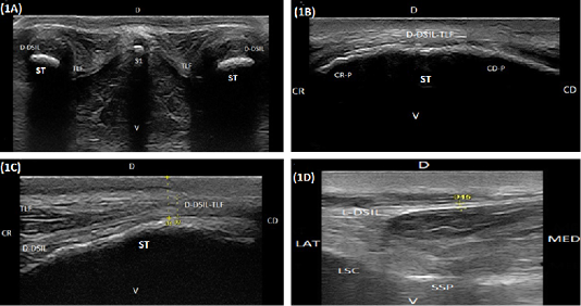
(A) Compound transverse sonogram (10 MHz linear tendon probe, 6 cm depth) at the dorsal aspect of the sacroiliac region of a 6-year-old cow showing sacral tuber (ST), the thoracolumbar fascia (TLF), the dorsal portion of the dorsal sacroiliac ligament (D-DSIL). first sacral spinous process (S1), dorsal direction (D) and ventral direction. (B) Compound longitudinal sonogram (14 MHz linear tendon probe, 5cm depth) at the dorsal area of sacral tuber of a 7- year- old cow showing sacral tuber (ST), the dorsal portion of the dorsal sacroiliac ligament- thoracolumbar fascia combination (D-DSIL-TLF), cranial margin of sacral tuber (CR-P), caudal margin of sacral tuber (CD.P), cranial direction (CR), caudal direction (CD), dorsal direction (D) and ventral direction (V). (C) Compound longitudinal sonogram at the dorsal area of sacral tuber of the same animal in Figure. 1B. showing the dorsal aspect of the dorsal sacroiliac ligament (D-DSIL), thoracolumbar fascia (TLF), sacral tuber (ST), cranial direction (CR), caudal direction (CD), dorsal direction (D) and ventral direction (V). (D) Transverse sonogram (10 MHz linear tendon probe, 6 cm depth) of an 8-year-old cow showing the lateral aspect of the dorsal sacroiliac ligament (L-DSIL), the lateral sacral crest (LSC), the sacral spinous process (SSP), lateral direction (LAT), medial direction (MED), dorsal direction (D) and ventral direction (V).(A) Compound transverse sonogram (10 MHz linear tendon probe, 6 cm depth) at the dorsal aspect of the sacroiliac region of a 6-year-old cow showing sacral tuber (ST), the thoracolumbar fascia (TLF), the dorsal portion of the dorsal sacroiliac ligament (D-DSIL). first sacral spinous process (S1), dorsal direction (D) and ventral direction. (B) Compound longitudinal sonogram (14 MHz linear tendon probe, 5cm depth) at the dorsal area of sacral tuber of a 7- year- old cow showing sacral tuber (ST), the dorsal portion of the dorsal sacroiliac ligament- thoracolumbar fascia combination (D-DSIL-TLF), cranial margin of sacral tuber (CR-P), caudal margin of sacral tuber (CD.P), cranial direction (CR), caudal direction (CD), dorsal direction (D) and ventral direction (V). (C) Compound longitudinal sonogram at the dorsal area of sacral tuber of the same animal in Figure. 1B. showing the dorsal aspect of the dorsal sacroiliac ligament (D-DSIL), thoracolumbar fascia (TLF), sacral tuber (ST), cranial direction (CR), caudal direction (CD), dorsal direction (D) and ventral direction (V). (D) Transverse sonogram (10 MHz linear tendon probe, 6 cm depth) of an 8-year-old cow showing the lateral aspect of the dorsal sacroiliac ligament (L-DSIL), the lateral sacral crest (LSC), the sacral spinous process (SSP), lateral direction (LAT), medial direction (MED), dorsal direction (D) and ventral direction (V).
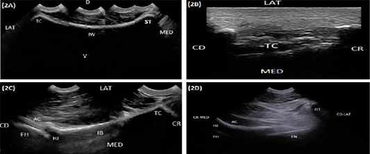
(A) Compound longitudinal sonogram (6.6 MHz micro convex probe, 10 cm depth) in a 5-year-old cow showing iliac wing (IW), sacral tuber (ST), tuber coxae (TC), lateral direction (LAT), medial direction (MED), dorsal direction (D) and ventral direction (V). (B) Longitudinal sonogram (10 MHz linear tendon probe, 5cm depth) in an 8-year-old cow showing tuber coxae (TC) lateral direction (LAT), medial direction (MED), cranial direction (CR) and caudal direction (CD). (C) Compound longitudinal sonogram (6.6 MHz micro convex probe, 10 cm depth) in a 5-year-old cow showing the iliac body (IB), hip joint (HJ), tuber coxae (TC), acetabulum (AC), femoral head (FH), lateral direction (LAT), medial direction (MED), cranial direction (CR) and caudal direction (CD). (D) Oblique sonogram (6.6 MHz micro convex probe, 15 cm depth) in an 8-year-old cow showing the hip joint (HJ), the acetabulum (AC), femoral head (FH), femoral neck (FN), greater trochanter (GT), craniomedial direction (CR-MED), and caudolateral direction (CD-LAT).
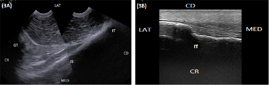
(A) Compound longitudinal sonogram (6.6 MHz micro convex probe, 11 cm depth) in a 6-year-old cow showing the ischium (IS), ischial tuber (IT), greater trochanter (GT). CR, Lateral direction (LAT), medial direction (MED), cranial direction (CR) and caudal direction (CD). (B) longitudinal sonogram (10 MHz linear tendon probe, 5 cm depth) in an 8-year-old cow showing ischial tuber (TI), lateral direction (LAT), medial direction (MED), cranial direction (CR) and caudal direction (CD).
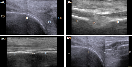
(A) Compound longitudinal sonogram (10 MHz linear rectal probe, 5cm depth) in a 7-year-old cow showing tabula of the ischium (TI), obturator foramen (OF), cranial direction (CR) and caudal direction (CD). (B) Transverse sonogram (10 MHz linear rectal probe, 5cm depth) of the caudal half of the pelvic floor in of the same animal in Figure: 4A showing the pelvic symphysis (PS) and tabula of the ischium (TI). (C) Transverse sonogram (10 MHz linear rectal probe, 5cm depth) of the cranial half of the pelvic floor of the same animal in Figure: 4A showing the pelvic symphysis (PS) and tabula of the ischium (TI). (D) Compound longitudinal sonogram (10 MHz linear rectal probe, 10cm depth) of the of the same animal in Figure 4A showing obturator foramen(OF), blood vessels (BL. V), nerve (N), tabula of the ischium (TI), pubis (P), caudal direction (CD) and cranial direction (CR).
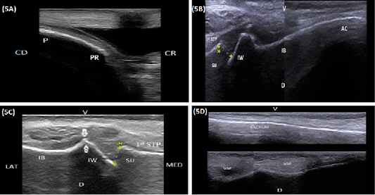
(A) Longitudinal sonogram (10 MHz linear rectal probe, 5 cm depth) of the pubis (P) in a 7-year-old cow showing the pubic rim (PR), caudal direction (CD) and cranial direction (CR). (B) Longitudinal sonogram (10 MHz linear rectal probe, 5 cm depth) in a 6-year-old cow showing the medial aspect of the acetabulum (AC), the medial aspect of the iliac body (IB), iliac wing (IW), sacroiliac joint (SIJ), first sacral transverse process (1st STP), ventral direction (V) and dorsal direction (D). (C) Longitudinal sonogram (10 MHz linear rectal probe, 7 cm depth) in a 5-year-old cow showing the sacroiliac joint (SIJ), iliac wing (IW), first sacral transverse process (1st STP), lateral direction (LAT) and medial direction (MED). Note: A prominent bony lip (arrows) was imaged at the point of junction between the iliac wing and body (IB). (D) Longitudinal sonogram (10 MHz linear rectal probe, 5 cm depth) of the same animal in Figure: 5C showing the ventral aspect of the sacrum (S), ventral sacral foramina (VSF), ventral direction (V) and dorsal direction (D). Proximal is the central portion and Distal is the lateral portion.


