Research Journal for Veterinary Practitioners
Case Report
Surgical Management of Malignant Histiocytosis in a Buffalo
Logaiyan SivaSudarshan, Jetty Devarathnam, Pidugu Amaravati, Gurram Kamalakar, Rajapantula Mahesh, Narpala Sumiran, Rayadurgam Venkata Suresh Kumar
College of Veterinary Science, Proddatur (A. P.), India.
Abstract | A case of malignant histiocytosis in a buffalo is described. A 4 year old Non–descript Female buffalo was presented with 1 year history of pedunculated, soft and ulcerated mass on the left lateral angle of ilium. Fine needle aspiration biopsy revealed malignancy. The mass was surgically treated under local anaesthesia. Based on histological findings it was diagnosed as malignant histiocytosis. After surgery the animal recovered without any complications and recurrence.
Keywords | Malignant tumour, Histiocytic sarcoma, Histiocytosis, Buffalo
Editor | Muhammad Abubakar, National Veterinary Laboratories, Islamabad, Pakistan.
Received | July 07, 2014; Revised | September 11, 2014; Accepted | September 13, 2014; Published | November 7, 2014
*Correspondence | Jetty Devarathnam, College of Veterinary Science, Proddatur (A. P.), India; Email: drvictory84@gmail.com
Citation | SivaSudarshan L, Devarathnam J, Amaravati P, Kamalakar G, Mahesh R, Sumiran N, Kumar RVS (2015). Surgical management of malignant histiocytosis in a buffalo. Res. J. Vet. Pract. 3 (1): 10-12.
DOI | http://dx.doi.org/10.14737/journal.rjvp/2015/3.1.10.12
ISSN | 2308–2798
Copyright © 2015 SivaSudarshan et al. This is an open access article distributed under the Creative Commons Attribution License, which permits unrestricted use, distribution, and reproduction in any medium, provided the original work is properly cited.
Malignant histiocytosis in domestic animals is a multisystemic neoplasm that proliferates primarily in the spleen, lungs or bone marrow. Secondary sites of neoplastic proliferation include lymph nodes and liver, with several other organs affected subsequently (Suzuki et al., 2003). It is the most destructive syndrome of all histiocytic neoplasms (Goldschmidt and Hendrick, 2002) and was reported in cattle (Anjiki et al., 2000), dogs (Moore et al., 2006), horses (Lester et al., 1993) and cats (Trost et al., 2008). The present case report describes one such rare finding of malignant histiocytosis in a female buffalo and its surgical management.
A 4 year old non–descript female buffalo was presented to the Teaching Veterinary Clinical Complex, Proddatur (Andhrapradesh, India) a history of soft and ulcerated swelling on the left lateral angle of ilium (Figure 1). It was reported that the mass showed a slow progressive growth over a period of one year and was ulcerated. On palpation the mass was soft and was attached to skin at the ilium. Base of the mass was pedunculated and broad. Fine needle aspiration cytology revealed malignancy. No other abnormal clinical manifestations were observed. The case was decided to treat surgically.
The buffalo was prepared for anesthesia and surgery by withholding water and feed for 18 h. The animal was sedated with xylazine hydrochloride (Xylaxin: Indian immunologicals) @ 0.1mG/kG body weight and lignocaine hydrochloride (Xylocaine: Astra Zeneca) was infiltrated around the incision site. The surgical site was aseptically prepared. An elliptical incision was made on the skin around the base of tumour mass. By blunt dissection, the tissues were separated. Bleeding points were controlled by ligating major blood vessels and the tumour mass was excised completely (Figure 2). A small piece of excised tumour mass was preserved in 10 % formalin and sent for histopathological examination. Sub–cuticular sutures were applied with chromic catgut No: 1 and the skin was opposed in horizontal mattress suture pattern using nylon suture material (Figure 3). Postoperatively Streptopenicillin @ 20mG/kG body weight and Meloxicam @ 0.3mG/kG were administered for 5 consecutive days followed by daily dressing of wound 1% povidone iodine solution. Gamma benzene hexachloride ointment was used as fly repellent. Skin sutures were removed on 10th day post–operative. The animal had uneventful recovery without any complications. No recurrence was reported after surgery for a period of six months.

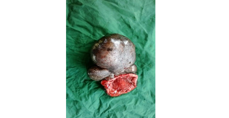
Figure 2: Excised tumor mass
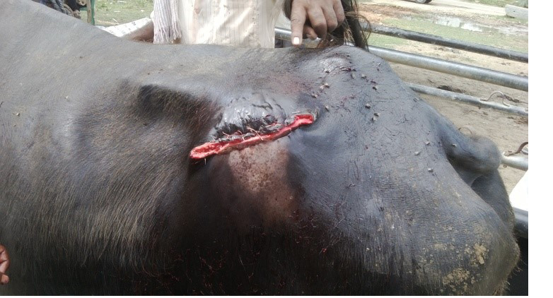
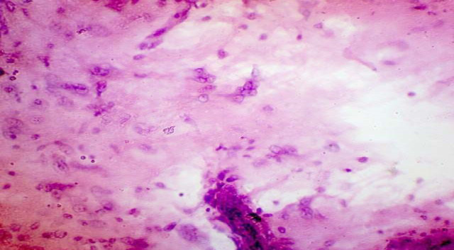
Figure 4: Photomicrograph of tumour mass showing large round to polygonal cells with round nuclei and abundant cytoplasm indicating histiocytes, along with few multinucleated forms (H & E)
Histological sections (H & E staining) of the tumour mass revealed multifocal, large round to polygonal cells with round nuclei and abundant cytoplasm indicating histiocytes, along with few multinucleated forms (Figure 4). Based on cytological and histopathological examination the tumour mass was diagnosed as malignant histiocytosis.
In the present case, histological sections of excised tissue mass from buffalo ilium showed histiocytes indicating that the condition might be one of the types of histiocytic neoplasms. Malignant histiocytosis is characterized by progressive and invasive multi–systemic proliferation of atypical histiocytes and their precursors (Moore et al., 2006). Animals with malignant histiocytosis have non–specific clinical signs (Trost et al., 2008). In the present case also the animal did not show any conspicuous clinical findings definitive of malignant histiocytosis. The definitive diagnosis of malignant histiocytosis is made by histopathological examination. Malignant histiocytosis is a diagnosis of exclusion and can be done when the tumour cells have morphological, immunohistochemical and ultrastructural characteristics with those of histiocytes (Freeman et al., 1995). It can be differentiated from other histiocytic diseases for presenting high levels of cellular atypia (Trost et al., 2008). In the present case, histological sections revealed abnormal shaped cells ie., cellular atypia with abundant cytoplasm resembling histiocytes indicating malignant histiocytosis. Localized neoplasms are best treated by surgical excision. In the present case lesions were limited to skin. Multisystemic involvement could not be diagnosed as the animal was clinically normal other than the growth at the ilium. Localized histiocytic sarcoma is treated by surgical excision (Fulmer and Mauldin, 2007). Therefore surgical excision was opted for management of the condition.
CONCLUSION
Malignant histiocytosis is a rare neoplastic condition observed in buffaloes. If the neoplasm is localized, surgical excision can be considered for treatment. In case of metastasis systemic chemotherapy is recommended. However malignant histiocytosis with metastasis has poor prognosis even after aggressive therapy.
REFERENCES