Journal of Animal Health and Production
Research Article
J. Anim. Health Prod. 5(1): 29-34
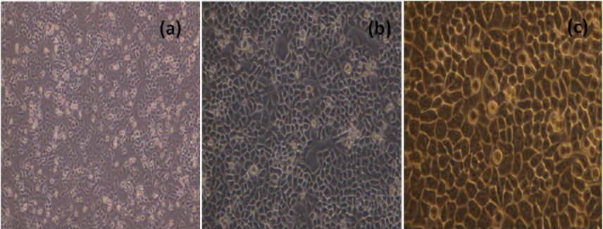
Figure 1
Uninfected MCF-7 cells 24 h(a)100X, 48h (b)200X and 72h (c) 400X; after subculture
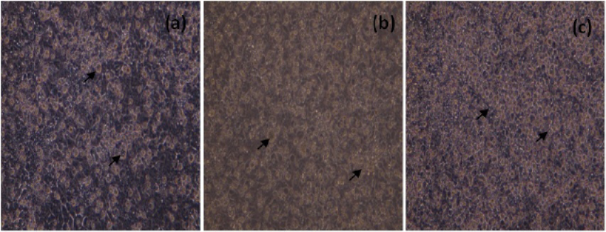
Figure 2
Unstained, MCF-7 cells infected with NDV (200X), 24h (a), 48h(b) and 72h (c) p.i. Arrows indicate, rounding of cells and detachment of monolayer
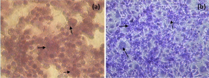
Figure 3
H and E (a) and Giemsa (b) staining of NDV infected MCF-7 cells (400X, Inverted microscope). Arrows indicate, rounding of cells, fusion of cell cytoplasm and syncytia formation


